-
Implications of starvation-induced change in right dorsal anterior cingulate volume in anorexia nervosa.
- McCormick LM, Keel PK, Brumm MC, Bowers W, Swayze V, Andersen A, Andreasen N.
- Int J Eat Disord. 2008 Nov;41(7):602-10. doi: 10.1002/eat.20549.
-
- PMID: 18473337
- Free PDF
- Free PMC Article
Comments from Dr. McCormick
Here are actual MRIs from an AN patient in one of my studies that demonstrates global brain atrophy before and restoration after weight gain. These correspond to one of my articles that shows a 15% decrease in right dorsal ACC volume with starvation, which was correlated to reduced PIQ often seen in patients with AN. The degree to which the right dorsal ACC volume reverted back to normal was related to outcome as well. I have also attached an actual MRIs from an AN patient in one of my studies that demonstrates global brain atrophy before and restoration after weight gain.


- Laurie McCormick, MD
- Assistant Professor
- University of Iowa Carver College of Medicine
- Department of Psychiatry, Psychiatric Iowa Neuroimaging Consortium
Comments from Dr. Bohon
The brain on the left is from an underweight AN patient. She has not yet recovered, though, so we do not have a weight-restored scan for her. It is probably the best one we have, though, to really get a visual sense of atrophy. Our other pre- post- scans are much more subtle and would really require volumetrics to get a sense of the change rather than just a visual glance. is the brain on the right is one of our control subjects as a comparison. Obviously, it's a different brain, so getting the slices to match isn't perfect, but you can at least get a comparison of underweight brain vs.normal weight brain - even if it's a different person.
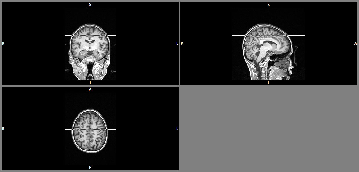
Underweight AN Patient
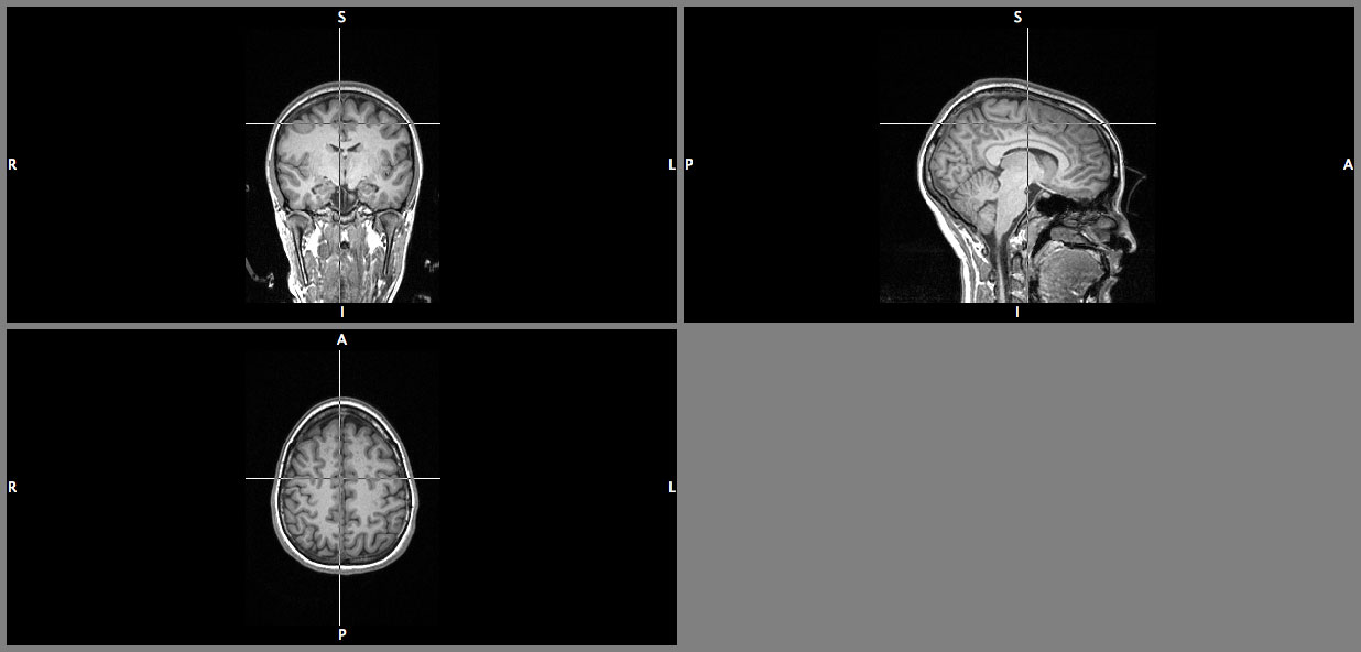
Normal weight brain
- Cara Bohon, Ph.D.
- Postdoctoral Scholar-Fellow
- University of California, Los Angeles
- Department of Psychiatry and Biobehavioral Sciences